
Fig. 6.
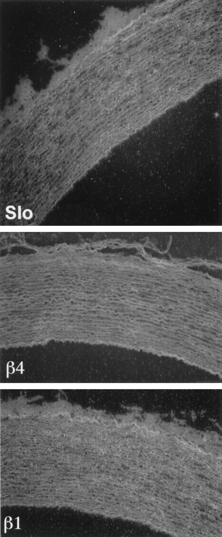
Analysis of Slowpoke α and β subunit expression in monkey aorta. Emulsion autoradiograms of sections of monkey aorta labeled with antisense probes to Slowpoke α (Slo, top panel), β4 (middle panel), and β1 (bottom panel) subunits were viewed under dark-field illumination. Silver grains representing mRNA encoding α and β1 subunits are readily visible over smooth muscle layers in the wall of the monkey aorta, whereas β4 expression is not detectable (10× magnification).