
Fig. 2.
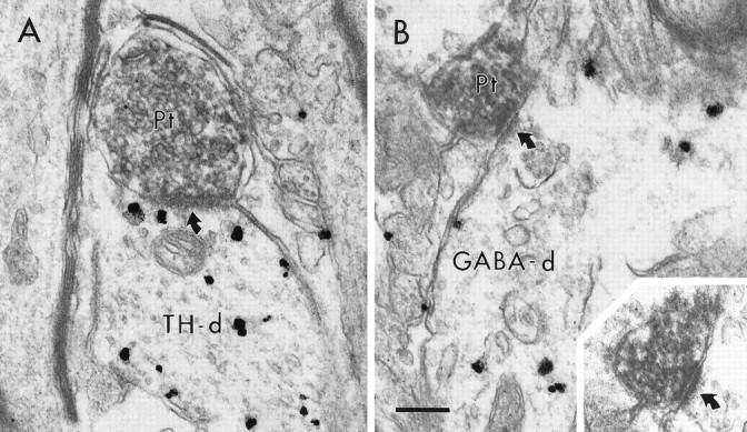
Electron micrographs showing synaptic contacts (curved arrows) of BDA-labeled PFC terminals (Pt) onto dendrites containing immunogold–silver labeling for TH (A, TH-d) or GABA (B, GABA-d). In B, the synapse formed by the Pt on the GABA-d is also prominent in an adjacent section (inset). Scale bar, 0.25 μm.