
Fig. 1.
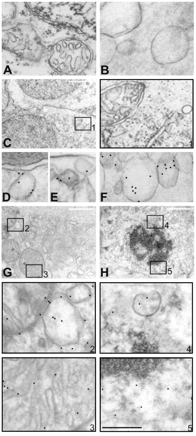
Internalization of hsp27 antibody by human retina observed by immunogold labeling and electron microscopy. No gold particles were observed within neuronal cells in control retina incubated without antibody (A) or incubated with control antibodies, anti-IgG (B), or anti-calbindin-D (C). C, Micrograph of retinal ganglion cells after incubation with calbindin-D antibody for 12 hr. Higher magnification micrograph of selected area inA is shown in box 1, which indicates negative gold labeling. However, after incubation with hsp27 antibody, intracellular gold particles were observed in a time-dependent pattern. Immunolabeling was positive in endosomes (D,E) and vesicles (F) of the retinal ganglion cells incubated with hsp27 antibody for 30 min. Micrographs of two retinal cells incubated with hsp27 antibody for 6 and 12 hr, respectively, are shown in G and H. Higher magnification micrographs of selected areas in G andH are shown in boxes with corresponding numbers.Boxes B2 and 3 show mitochondrion and vesicules, and boxes 4 and 5 show perinuclear area. After incubation with hsp27 antibody for 6 or 12 hr, gold particles were mostly observed in vesicular structures of various size and mitochondria (G, H). In addition, perinuclear areas and nuclei of the retinal cells incubated with hsp27 antibody exhibited immunogold labeling (H). Notice the double membrane and internal cristae of mitochondria in G and condensed nuclear chromatin in H. Black scale bar:A, B, D–F, 0.5 μm; white scale bar: C, G,H, 2 μm.