
Fig. 5.
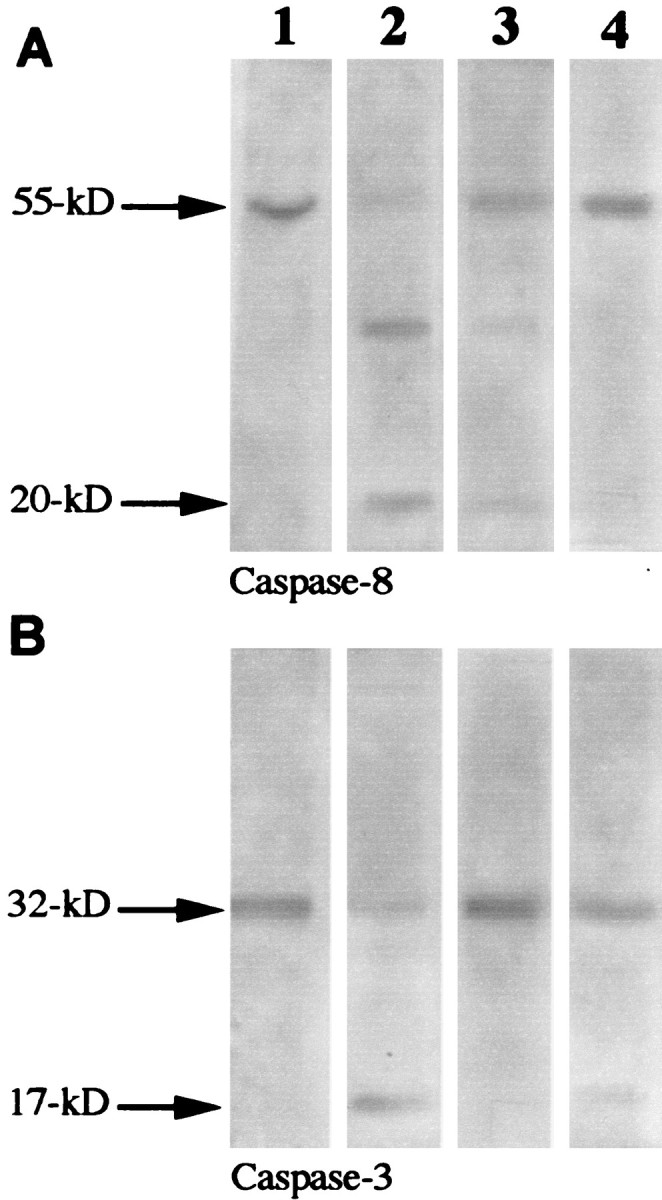
Western blot findings demonstrating caspase cleavage: column 1, control retinal cells (E1A.NR3);column 2, retinal cells incubated with hsp27 antibody (100 μg/ml) for 24 hr; column 3, retinal cells incubated with hsp27 antibody in the presence of a nonselective caspase inhibitor, B-D-FMK (50 μm); column 4, retinal cells incubated with hsp27 antibody in the presence of the caspase-8 inhibitor Z-IETD-FMK (20 μm). Although no cleavage of caspase-8 (A, column 1) or caspase-3 (B, column 1) was detected using the lysates of the control retinal cells, cleavage of caspase-8 and caspase-3 was observed using retinal cells incubated with hsp27 antibody. A 55 kDa immunoreactive band corresponding to caspase-8 cleaved to 30 and 20 kDa products is shown in A,column B. A 32 kDa pro-enzyme caspase-3 cleaved to a 17 kDa subunit is shown in B, column 2. Incubation of the retinal cells with hsp27 antibody in the presence of caspase inhibitors prevented specific caspase cleavage. B-D-FMK effectively inhibited caspase-3 and partially prevented caspase-8 cleavage (column 3); Z-IETD-FMK inhibited the cleavage of caspase-8 and only partially inhibited the cleavage of caspase-3 (column 4).