
Fig. 5.
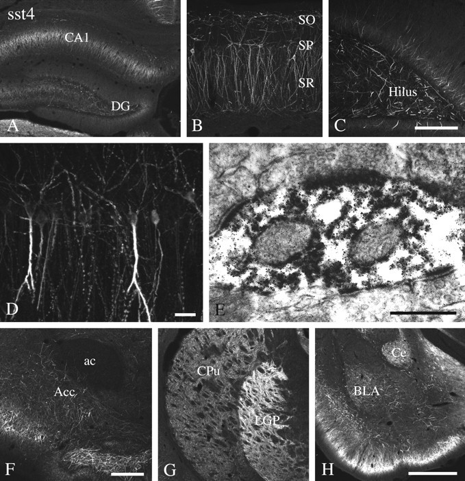
Immunofluorescent and electron micrographs showing the regional and subcellular localization of sst4-Li in rat forebrain. A–D, F–H, Coronal rat brain section immunofluorescently stained with affinity-purified anti-sst4 antibodies (6002). E, Rat brain sections from the hippocampal CA1 region were processed for immunoperoxidase detection of the anti-sst4 antibody. Note that sst4-Li is enriched in the hippocampal formation with high levels found in the Ammon's horn and the hilar region of the dentate gyrus. sst4-Li was also distributed along neuronal processes in the nucleus accumbens, striatum, and amygdala. At the electron microscopic level, immunoperoxidase product was always postsynaptic, and some instances of immunolabeling at asymmetrical synapses were found in the hippocampal CA1 region (E). ac, Anterior commissure;Acc, nucleus accumbens, BLA, basolateral amygdaloid nucleus; Ce, central amygdaloid nucleus,CPu, caudate-putamen; DG, dentate gyrus;LGP, lateral globus pallidus; SO, stratum oriens; SP, stratum pyramidale, SR, stratum radiatum. Scale bars: A, G,H, 250 μm; B, C, 50 μm; D, 10 μm; E, 0.2 μm;F, 100 μm.