
Fig. 1.
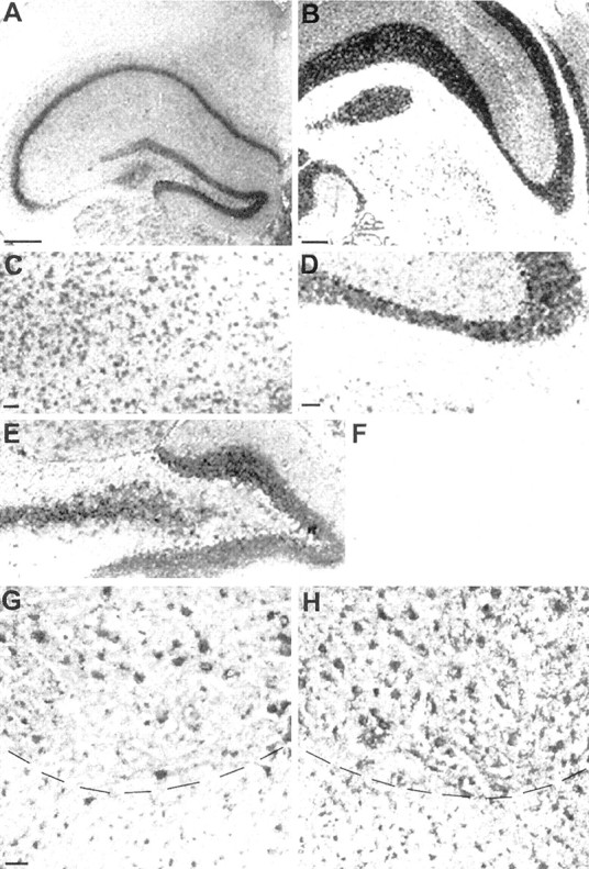
ADAM8 mRNA in the CNS of normal mice as shown byin situ hybridization (coronal and transversal sections; dorsal is up). A, Cerebral cortex, anterior part with hippocampus (lateral is left).B, Brainstem with part of cerebellum (lateral isright). C, Cortical layer (detail ofA). D, Granular layer of the cerebellum with Purkinje cells (detail of B). E, Gyrus dentatus (detail of A). F, Negative control (sense probe), detail of cerebellum, as in D.G, H, Cervical spinal cord, ventral part; comparison between wild type (G) and wobbler (H). Dashed lines indicate borders between gray (top) and white (bottom) substance. Scale bars: A,B, 500 μm; (in C) C,E, 100 μm; (in D) D,F, 100 μm; (in G) G,H, 50 μm.