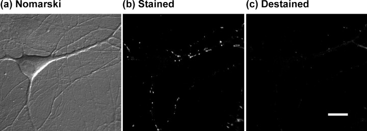
Fig. 1.
Example of FM 1-43 staining.a, Nomarski image of a pyramidal neuron in a mixed neuronal–glial dissociated culture. b, Fluorescence image of the same field acquired after FM 1-43 staining, using a 100-stimulus train at 10 Hz and a 40 sec exposure to FM 1-43. Discrete fluorescent puncta are visible, often at sites corresponding to dendritic interactions visible in the Nomarski image. c, Fluorescence image acquired after destaining using a 900-stimulus train at 10 Hz. Note that the punctate staining is primarily absent, leaving a dim background attributable to nonspecific membranous staining by FM 1-43. Scale bar, 10 μm.

