
Fig. 4.
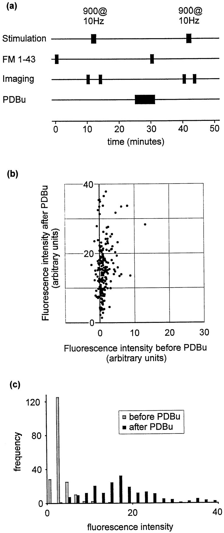
Strong effect of PDBu on spontaneous release.a, Protocol used to measure spontaneous release occurring during 1 min. FM 1-43 was applied for 1 min in the absence of stimulation. As in other protocols, destaining was performed using a 900-stimulus train at 10 Hz. For these experiments a third trial was also performed in which vesicles were stained using a 100-stimulus train at 10 Hz (trial not shown). The data from this third trial were used verify that the sites of fluorescence staining were positionally stable. This was necessary during these experiments, because many puncta exhibited very weak loading during the first trial, raising the possibility that a fluorescent punctum that was visible only during the second trial represented mobile fluorescence rather than a site at which recycling had been promoted by PDBu treatment. This precaution should therefore exclude the possibility that our selection procedure favored PDBu-sensitive synapses. b, Scatter plot showing fluorescence intensities before and after PDBu treatment.c, Data presented as a frequency histogram.