Fig. 3.

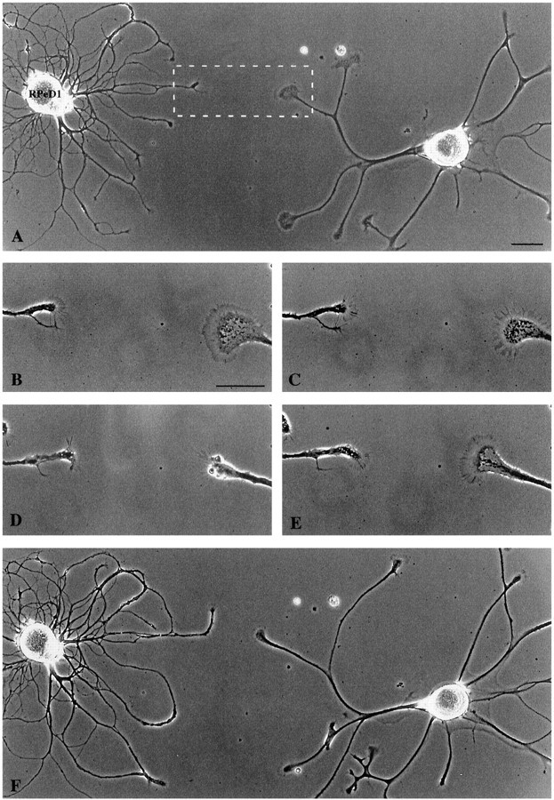
RPeD1 growth cones induced the collapse of a nontarget cell growth cone. A, RPeD1 and a nontarget cell were plated in close proximity and extended neurites over a 24 hr period. Scale bar, 75 μm. The boxed area represents the magnified images in B–E. The nontarget growth cone approached within ∼60 μm of the RPeD1 growth (B, 15 min) and collapsed (C, 30 min). Scale bar, 30 μm. The nontarget cell growth cone was fully collapsed at 60 min (D) and recovered over the next 45 min (E). F, Lower magnification demonstrating that the nontarget growth cone did not contact RPeD1.
