
Fig. 5.
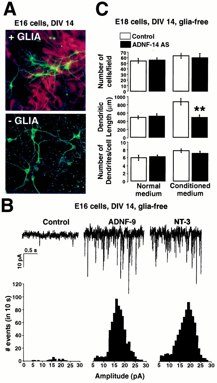
Physiological and morphological effects of exogenous and endogenous ADNF on glia-free neuronal cultures. To produce glia-free neuronal cultures, cells derived from hippocampus at E16 (A, B) and E18 (C) were plated on coated glass coverslips and grown for 2 weeks in the presence of the antimitotic agent floxuridine to prevent astrocytic growth.A, Neurons grown on glial monolayers or directly on glass were stained with the neuronal marker MAP2ab (green) and the glial marker GFAP (red). No GFAP staining was detected in glia-free cultures. B, Top trace, Spontaneous activity recorded from E16 glia-free cultures either untreated or treated for 2 weeks with 10−8m ADNF-9 or 20 ng/ml NT-3. Bottom histogram, Distribution of the frequency of mPSCs amplitudes. The frequency of the mPSCs was markedly enhanced in cultures treated with ADNF-9 or NT-3 compared with untreated cultures. C, Effect of ADNF-14 AS treatment on numbers and morphology of neurons in E18 glia-free cultures. Cultures were grown for 2 weeks in glia-derived conditioned medium (Conditioned medium) or fresh medium (Normal medium). CM markedly induced dendritic growth, an effect that was blocked by co-treatment with ADNF-14 AS. Treatment with ADNF-14 AS did not have any influence on the morphology of neurons grown in the absence of CM. The number of cells was comparable in all culture conditions. Results are the mean ± SEM of data from 10 different fields in a single culture experiment and are representative of three such experiments. **p < 0.01, t test. DIV, Days in vitro.