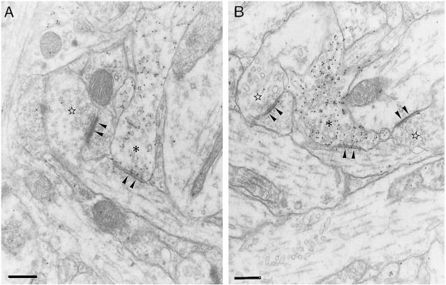
Fig. 8.
Electron photomicrographs of GABAergic synapses in control (A) and bicuculline-treated (B) slices. The asterisks mark labeled presynaptic terminals of GABAergic synapses, which contain pleomorphic synaptic vesicles, and establish type II synaptic complexes. Note the difference in the densities at the cytoplasmic faces of the postsynaptic differentiations (arrowheads) in GABAergic and non-GABAergic (stars) synapses. The latter correspond to type I synaptic complexes. Scale bars, 300 nm.

