
Fig. 7.
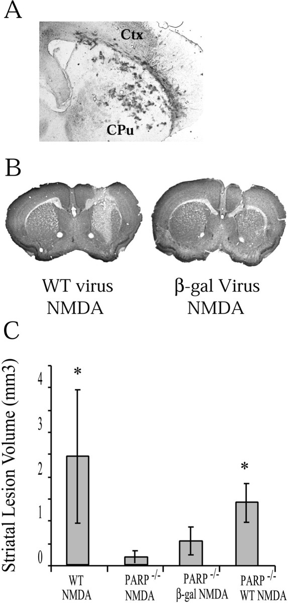
A, Coronal section through striatum 4 d after intrastriatal injection of Sindbis virus-expressinglacZ. Virus is prominent in part of the striatum. These results were replicated at least three times. B, Coronal section of a PARP-1−/− mouse receiving Sindbis virus-expressing cDNA of WT PARP-1 and then 48 hr after an intrastriatal injection of 20 nmol of NMDA. The section is stained with cresyl violet and demonstrates a demarcated area of NMDA damage.C, Lesion volumes (mean ± SD) of PARP-1−/− receiving Sindbis virus encoding PARP-1 WT cDNA (n = 3) demonstrate lesion volumes approaching that of WT mice receiving NMDA. PARP-1−/− mice receiving Sindbis virus only encoding lacZ (n = 3) demonstrate significantly lower lesion volumes (significance determined by ANOVA with Newman–Keuls post hoc analysis; *p < 0.05, significantly different from PARP-1−/− NMDA).