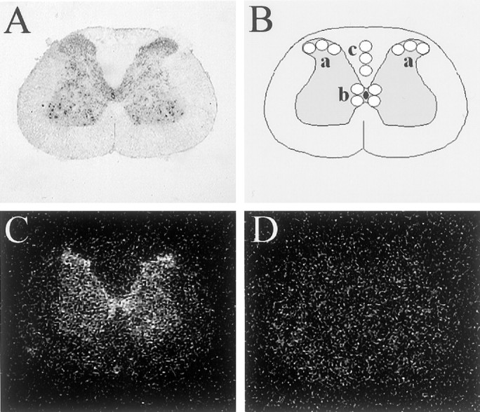
Fig. 1.

125I-NDP-MSH binding to rat spinal cord sections. A, Nissl staining of a representative spinal cord section, demonstrating the neuroanatomy.B, Diagram representing the sampling template used for determining 125I-NDP-MSH binding in x-ray film autoradiograms of rat spinal cord sections. a, Superficial dorsal horn (left and right); b, lamina X;c, dorsal white matter column used for determining background value. For each region, the mean value of three (superficial dorsal horn and background) or four (lamina X) samples was calculated.C, D, x-Ray film autoradiogram of125I-NDP-MSH binding to a representative rat spinal cord section. Sections were incubated with 125I-NDP-MSH in the absence (C) or presence (D) of 3 μm non-iodinated NDP-MSH. Specificity of binding present in C is demonstrated by its inhibition inD.
