Fig. 6.

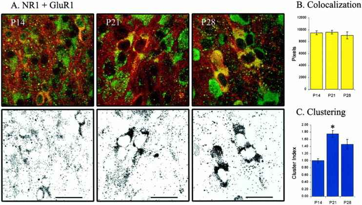
AMPA and NMDA receptor subunit colocalization is stable over ON/OFF sublamination. A, NR1 (NMDA) and GluR1 (AMPA) subunit expression and colocalization at P14 (n = 15 images from 2 animals), P21 (n = 22 images from 4 animals), and P28 (n = 14 images from 3 animals). Top row shows examples of labeling of NR1 (green) and GluR1 (red) through ON/OFF sublamination. Colocalized labeling is shown inyellow. Bottom row shows only the colocalized labeling from the corresponding row above.B, Colocalization remains constant from P14 to P28 (p > 0.05). C, The clustering of colocalized pixels is similar before and after sublamination (p > 0.05), although it increases during the third postnatal week (p< 0.05). Scale bar (in this and all subsequent figures), 25 μm.
