Fig. 8.
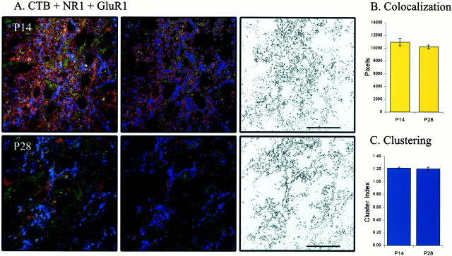
The pattern of AMPA–NMDA colocalization opposite retinogeniculate terminals is not different from the global pattern of colocalization in the LGN. A, NR1 (green) and GluR1 (red) labeling opposite CTB-labeled retinogeniculate terminals (blue).Left column shows examples of triple labeling at P14 and P28. Middle column shows pixels just opposite CTB labeling (for details, see Materials and Methods). Right column shows only the colocalized labeling at pixels derived from the corresponding column to theleft. B, Colocalization at P28 (n = 5 images from 1 animal) is unchanged from P14 (n = 8 images from 1 animal; p> 0.05). C, Likewise, the clustering of colocalized pixels is similar before and after sublamination (p > 0.05).

