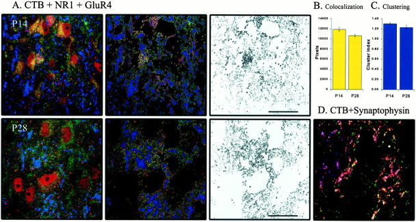
Fig. 9.
The pattern of AMPA–NMDA colocalization opposite retinogeniculate terminals is not unique to GluR1. A–C, Same convention as in Figure 8. GluR4–NR1 colocalization at P28 (n = 7 images from 1 animal) is unchanged from P14 (n = 3 images from 1 animal; p> 0.05). Clustering of colocalized pixels opposite retinogeniculate terminals is stable across ON/OFF sublamination (p > 0.05). D, CTB label overwhelmingly represents synaptic boutons, marked by synaptophysin, rather than fibers of passage. The vast majority of CTB label (blue) is colocalized with synaptophysin (green); colocalized label is shown inpink. There is also a significant amount of synaptophysin labeling independent of CTB, reflecting the nonretinal innervation of the LGN.

