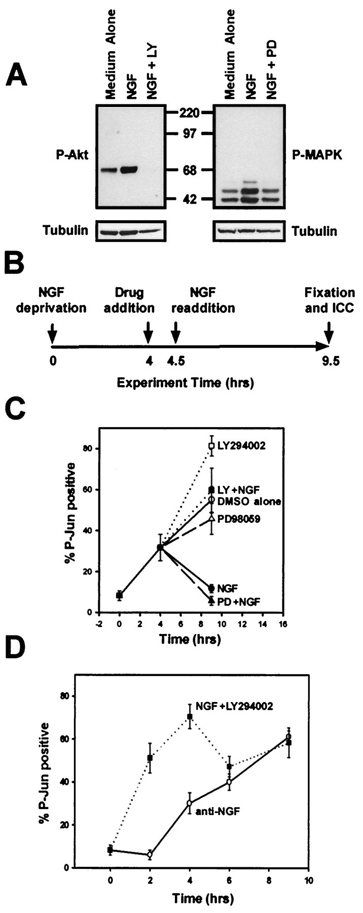
Fig. 1.

PI-3-K inhibits a cell death event proximal to c-Jun phosphorylation. A, Cultures of sympathetic neurons maintained in NGF (50 ng/ml) were treated with medium containing LY294002 (50 μm, left panel) or PD98059 (25 μm, right panel) to inhibit PI-3-K or MEK activities, respectively, or medium containing vehicle alone (DMSO, 0.1%) for 30 min. The cultures were then treated with medium alone, medium containing NGF (50 ng/ml), or medium containing NGF (50 ng/ml) in the continued presence of LY294002 or PD98059 for an additional 30 min. Detergent extracts were prepared, and equal amounts of the extracts were subjected to phospho-Akt (P-Akt) or phospho-MAPK (P-MAPK) immunoblotting. Reprobing the same blot with anti-α-tubulin after stripping demonstrates equal amounts of protein in the extracts. B, This schematic diagram depicts the experimental paradigm for the experiment described in C. Sympathetic neurons (5 DIV) were deprived of NGF for 4 hr, preincubated with medium containing LY294003 (50 μm), PD98059 (25 μm), or vehicle alone for 30 min, and next treated with medium containing NGF (300 ng/nl) in the continued presence of LY294002 or PD98059 for an additional 5 hr. C, Sympathetic neurons treated as in B were fixed after the treatments, and P-Jun was detected immunocytochemically. The number of neurons showing nuclear P-Jun staining is shown as a percentage of total cells. This experiment was performed in duplicate in three independent cultures.D, Sympathetic neurons were deprived of NGF (anti-NGF) or treated with LY294003 (50 μm) in the continued presence of NGF (NGF +LY29400). The cultures were fixed 2, 4, 6, or 9 hr after treatment and subjected to P-Jun immunocytochemistry. This experiment was performed in duplicate in three independent cultures. Error bars represent SEM.
