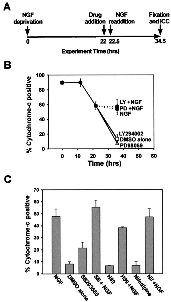
Fig. 3.

Neither PI-3-K nor MEK/MAPK mediates NGF-dependent inhibition of mitochondrial cytochrome c release.A, This schematic diagram represents the experimental paradigm used in B and C. Sympathetic neurons were deprived of NGF for 22 hr and next treated with vehicle alone (DMSO, 0.1%), LY294002 (50 μm), or PD98059 (25 μm) for 30 min. The cultures were then treated with medium alone or medium containing NGF (300 ng/ml) in the continued presence of LY294002 or PD98059 for an additional 12 hr before fixation. B, Cultures treated as in Awere immunostained for cytochrome c, and the number of neurons displaying punctate cytochrome c are shown as the percentage of total cells. This experiment was performed in duplicate in three independent cultures. Error bars represent SEM.Open symbols indicate cultures that were treated with medium alone in the presence of the listed pharmacological agent;closed symbols represent cultures treated with NGF.C, Sympathetic neuronal cultures were treated as inA, but the pharmacological agents added were the p38MAPK inhibitor SB203580 (30 μm), the PKA inhibitor H89 (5 μm), the L-type calcium channel antagonist nifedipine (200 nm), or vehicle alone, before NGF readdition. The number of neurons displaying punctate cytochrome c is displayed as a vertical bar chart depicting only the percentage of cytochrome c-positive cells remaining at the end of the treatments. This experiment was performed in duplicate in three independent cultures, with error bars representing SEM.
