
Fig. 4.
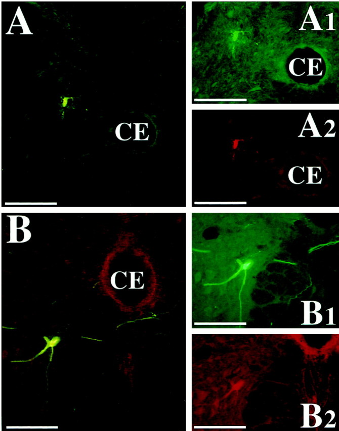
Photomicrographs of presumed spinal cord interneurons infected 4.5 and 5 d after PRV-Bablu injection into the diaphragm and PRV-152 injection into rectus abdominis. These neurons were dually immunostained with the red CY3 and the green CY2 fluorochromes, indicating that they made synaptic connections with both phrenic and abdominal motoneurons. The cells were located in lamina VII of the T8 (A) and lamina VIII of the L2 (B) spinal cord segments, ipsilateral to the injections. A1–B2 show the cell under illumination that excites one of the two fluorophors, whereasA and B were photographed under illumination that excites both fluorophors. Scale bars, 200 μm.