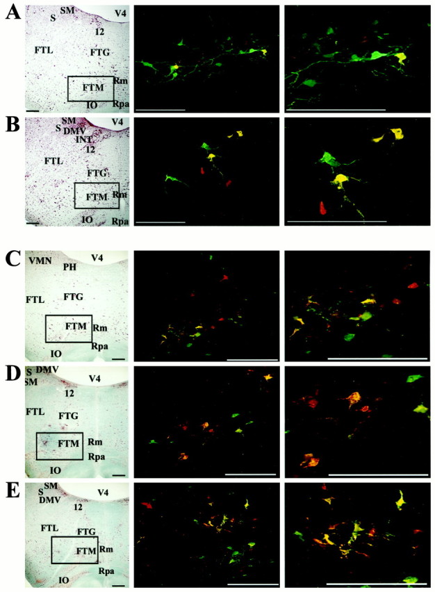
Fig. 6.

Location of premotor neurons in the MRF infected after injection of PRV into the diaphragm and rectus abdominis.A and B show infected neurons after a 4.5 d survival time, whereas C–E illustrate infection after a 5 d survival. In addition, A andD show labeling at ∼2.5 mm rostral to the obex, whereas B and E show labeling at ∼2.0 mm rostral to the obex, and C illustrates labeling at ∼3.0 mm rostral to the obex. Left column, Photomicrographs of brainstem sections (on the side ipsilateral to the injections) stained with a modified Kluver-Barrera method.Boxes surround the area containing infected neurons illustrated in the photomicrographs immediately on theright. Middle and right columns, Photomicrographs of infected neurons observed in the MRF. Red cells were only infected by retrograde transynaptic passage of PRV-Bablu from the diaphragm, green cells were only infected by retrograde transynaptic passage of PRV-152 from rectus abdominis, and yellow cells contain both viruses. Photomicrographs in the right column are a magnification of those in the middle column. Note that after a 5 d survival time, labeling is more prevalent and extends further rostrally than after a 4.5 d survival. Scale bars, 400 μm. Abbreviations are the same as in Figure 4, with the following additions: FTG, gigantocellular tegmental field;FTM, magnocellular tegmental field; INT, nucleus intercalatus; Rm, nucleus raphe magnus;Rpa, nucleus raphe pallidus.
