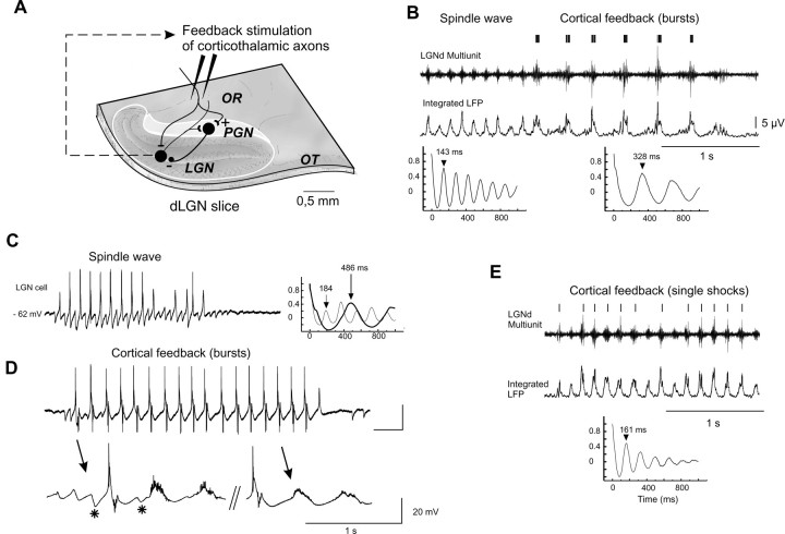
Fig. 2.
Control of thalamic oscillations by corticothalamic feedback in ferret thalamic slices. A,In the self-generating spindling thalamic slice, lateral geniculate (LGN) relay neuron axons projecting to the cortex via the optic radiation (OR) give off a collateral branch to the perigeniculate nucleus (PGN). The GABAergic PGN neurons generate direct inhibitory feedback to the relay neurons of the LGN. Corticothalamic axons run in the OR and synapse on LGN and PGN cells. Bipolar stimulating electrodes were placed in the OR. OT, Optic tract. B, A 7 Hz control spindle is slowed down to a 3 Hz oscillation by the feedback stimulation of OR at a latency of 20 msec after the detection of multiunit bursts activity (5 shocks; 100 Hz). Middle trace, Smooth integration of the multiunit signal (integrated local field potential). Bottom traces, Autocorrelation functions applied on the integrated LFP before and after the transition. C, Intracellular recording of a thalamocortical cell during spontaneous spindle oscillation. The first peak (184 msec) of the autocorrelation function indicates the period of network oscillation (i.e., the inverse of its beating frequency).D, Cortical feedback stimulations (4 shocks; 100 Hz; 50 msec delay) triggered by the burst firing of the cell slows the network oscillation to ∼2 Hz. Downward deflections in the cell are stimulation artifacts. Action potentials were truncated for clarity. The spike-triggering average below was made from before and after the first 2 sec of 12 and 64 oscillatory sequences, respectively; it indicates the persistence of fast compound IPSPs at the beginning of the oscillation (asterisk). An autocorrelation function of this slow oscillation is superimposed in C as thethick trace (486 msec). E, Weak (single shock) feedback stimulation delivered to OR.

