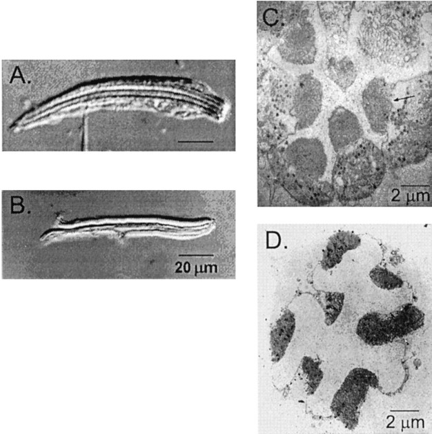
Fig. 1.
A, B, Light micrographs with Nomarski differential interference optics show an intact ommatidium (A) and an isolated rhabdomeral membrane (B) under standard recording conditions. A recording pipette is visible at the bottom ofA, attached to the plasma membrane of a single cell within the intact ommatidium. In the isolated rhabdomeral membranes shown in B, the plasma membranes normally present in intact cells have been stripped away. C, D, Transmitting electron micrographs show cross sections of an intact ommatidium (C) and an isolated rhabdomeral membrane (D). The electron-dense regions in the center of the intact ommatidium are the rhabdomeral membranes.

