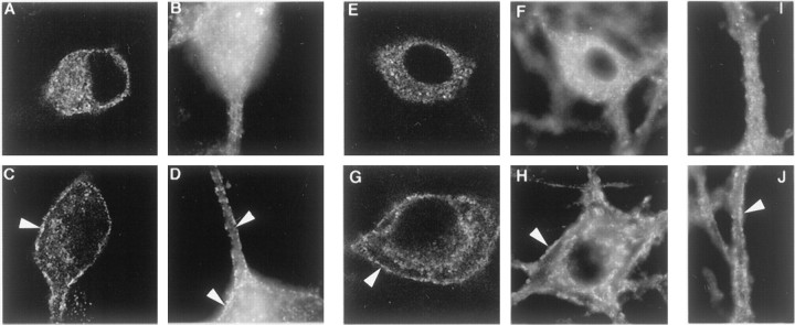
Fig. 6.
BDNF induced an increase in AP2 and clathrin at the surface of hippocampal neurons. To show whether NT treatment induced an increase in AP2 and clathrin associated with surface membranes in primary neurons, we used BDNF to treat rat hippocampal neurons. The distribution of AP2 was assessed by confocal (A, C) and epifluorescence (B, D) microscopy of neurons immunostained with AP.6. Clathrin distribution was assessed with confocal (E, G) and epifluorescence (F, H–J) microscopy of neurons immunostained for CHC with X22. BDNF (2 nm;C, D, G, H, J) or vehicle (A, B, E, F, I) was applied to cultured neurons for 2 min at 37°C before they were chilled, fixed, and processed for immunostaining. BDNF increased staining for AP2 (C, D) and CHC (G, H) at the plasma membrane (arrowheads). I and J show sections of neuronal processes and indicate that the BDNF effect also was registered here (J). The width of all panels is 55 μm.

