
Fig. 2.
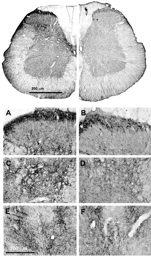
Immunohistochemical analysis of prodynorphin in lumbar spinal sections. Rats have been infused with either saline (control) or DAMGO for 7 d. Lumbar spinal cross sections (40 μm) were labeled with the antiserum for prodynorphin (1:40,000) and processed for DAB staining by the ABC method. Spinal cord halves are represented in close juxtaposition to allow visual comparison.Top, The lefthalfis obtained from the saline-infused control, and therighthalf is obtained from the DAMGO-infused rat. The micrographs were acquired via a Hamamatsu digital-imaging system with a Nikon microscope. Bottom, As seen under higher magnification, the superficial laminae (A, B), discrete cell bodies, and numerous fibers were labeled, whereas in laminae V and VI (C, D) and in lamina X (E, F), prodynorphin-IR was predominantly associated with fibers. Lumbar spinal sections from DAMGO-infused (B, D,F) rats exhibited a reduction of prodynorphin-IR in all the laminae stated above when compared with the saline-infused control (A, C, E). This reduction appears to be primarily caused by a loss of fiber staining in these laminae. In the superficial laminae, staining was still clearly present in cell bodies; the density of immunolabeled cell bodies was similar between control and DAMGO-infused rats.