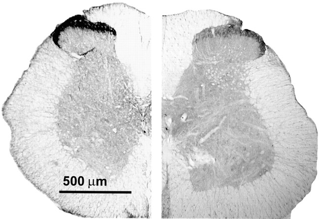
Fig. 3.
Immunohistochemical analysis of the μ-opioid receptor in lumbar spinal sections. Rats have been infused with either saline (control) or DAMGO for 7 d. Lumbar spinal cross sections (40 μm) were labeled with the antiserum for rat MOR (1:20,000) and processed for DAB staining by the ABC method. Spinal cord halves are represented in close juxtaposition to allow visual comparison. Thelefthalf is obtained from the saline-infused control, and the righthalf is obtained from the DAMGO-infused rat. Substantially higher MOR immunoreactivity is observed in the superficial laminae (I and II) of the control cord, indicating a greater concentration of μ-opioid receptors, than in that of the DAMGO-treated group. The micrographs were acquired via a Hamamatsu digital-imaging system with a Nikon microscope.

