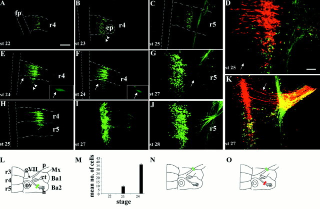
Fig. 2.
Retrograde labeling of motor neurons in r4 and r5 over a series of developmental stages. A–K, Ventral views of flat-mounted hindbrains showing retrograde labeling of facial BM and/or VM neurons. A, B, E, F, H, I, L, M, Labeling of BM neurons after fluorescein-dextran fills of the hyoid nerve (L, greenbar) at st. 22 (A), st. 23 (B), st. 24 (E, F), st. 25 (H), and st. 27 (I). Between three and five embryos were labeled at each stage. Note the presence of BM neurons in r5 from stage 23 onward. Many of the BM neurons in r5 occupy lateral positions (B, E, arrowheads), whereas others lie closer to the floor plate in a medial position (E, F, arrows). Higher power views of these medially positioned BM neurons are shown in the insets in E andF. C, G, J, Labeling of VM neurons after fluorescein-dextran fills of the palatine nerve (N, green bar) at st. 25 (C), st. 27 (G), and st. 28 (J). Four or five embryos were labeled at each stage. Note the curved trajectory of VM axons (G, arrow). D, K,Double-labeling of BM and VM subpopulations after fluorescein-dextran fills of the palatine nerve (O, green bar) and rhodamine-dextran fills of the hyoid nerve (O, red bar) at st. 25 (D) (n = 3) and st. 27 (K) (n = 5).D, Within r5 a single BM neuron in a medial position has been labeled (arrow), but the majority of facial motor neurons in r5 are located laterally. K, Motor neuron cell bodies at st. 27 occupy the same mediolateral position. The straight or slightly curved axon trajectories of r4 BM neurons are clearly seen (arrow). Presence of apparently double-labeled cells (yellow) in Dand K is an artifact caused by the superposition of labeled neurons at different depths when generating the confocalz-series shown here. Double-labeled cells were never observed in any single focal plane at all depths (data not shown).L, N, O, Schematics of ventral aspect of facial nerve and hindbrain indicating dextran labeling, and quantification of the mean (+SEM) number of BM neurons labeled in r5 at st. 22, 23, and 24 (M). Scale bar (in A):A, B, C, E, F, H, 100 μm. Scale bar (inD): G, I–K, 50 μm. fp,Floor plate; ep, exit point; gVII,geniculate ganglion; ov, otic vesicle; p,palatine nerve; ct, chorda tympani; h,hyoid nerve; Mx, maxilla; Ba1, first branchial arch; Ba2, second branchial arch.Dashed lines in A–C, E, F, andH indicate margins of floor plate and rhombomere boundaries.

