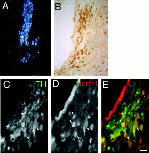
Fig. 1.
Localization of hcrtr1 in the LC.A, Dark-field photomicrograph of the LC hybridized with an hcrtr1 riboprobe. B, Immunocytochemical detection of hcrtr1. Note that most cells in the LC are labeled with the antibody. Scale bar, 50 μm. C–E, Immunofluorescence micrographs showing overlap (yellow, arrows) between hcrtr1-positive (red) and TH-immunoreactive (green) neurons in the LC region. Scale bar, 25 μm.

