
Fig. 3.
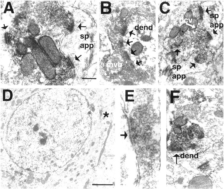
Electron micrographs taken from the shell of the nucleus accumbens in VCM+ (A–C), VCM− (D, E), and VEH-treated (F) rats. A, The dynorphin-immunoreactive terminal contacts two spines (arrows point to the active zone); one contact (rightside) is perforated. B, A dynorphin-immunoreactive ending forms a perforated, asymmetrical synapse (4 perforations labeled witharrows) with a dendritic shaft (dend). Note the multivesicular body (mvb) within the terminal. C, A dense core vesicle (dcv) is marked with awhitearrow. D, A dynorphin-positive terminal (asterisk) makes a symmetrical contact with the soma in a VCM− rat. Note the round, nonindented nuclear envelope, indicating that this is a medium spiny neuron. E, terminal seen in D (asterisk).F, A dynorphin-positive bouton makes a symmetrical synaptic contact with a dendritic shaft in a VEH-treated animal. Scale bars: A, 0.5 μm (valid for B, C, andF); E, 0.1 μm; D, 2 μm. sp app, spine apparatus.