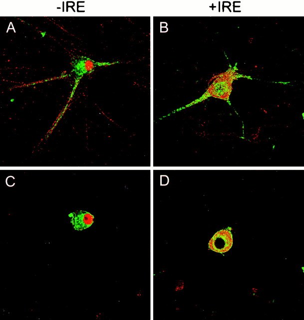
Fig. 8.
Confocal fluorescence micrographs of cultured rat sympathetic neurons treated for 5 d with BMP7 immunostained with antibodies specific for the −IRE (A, C) and +IRE (B, D) species of DMT1 and TfR.Green and red denote TfR and DMT1, respectively. Orange and yellow patchesrepresent areas in which TfR and DMT1 are colocalized. In the two optical sections (A, B), near the substratum on which the cells were grown, neurites are visible; inC and D, which are well above the substratum, staining patterns of the nucleus are most visible.

