
Fig. 1.
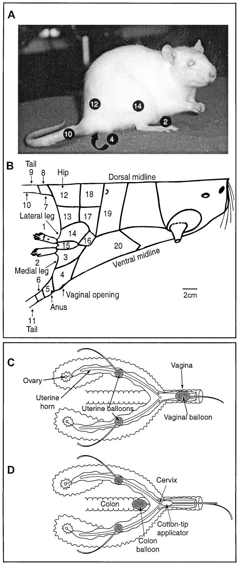
Stimulation protocol. B is a diagram of the rat's body traced directly from one half of a rat's pelt. Areas stimulated during the experiment are demarcated and numbered. Some of these are shown on the picture of the rat inA. C and D are diagrams of the female rat's reproductive tract. C shows the positions of the stimulating balloons implanted in the uterine horns and temporarily placed in the vaginal canal. D shows the position of the stimulating balloon temporarily inserted in the colon and the lubricated cotton swab applicator temporarily positioned next to the cervix.