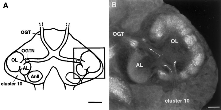
Fig. 1.
Morphology of the lobster deutocerebrum during late embryonic development. A, Schematic diagram outlining the arrangement of the deutocerebral neuropils that have been examined in previous serotonin depletion experiments (other neuropil areas in the deutocerebrum not shown). In decapod crustaceans, the OL, AL, andOGTN are innervated by branches of the olfactory projection neurons whose cell bodies are located lateral to the neuropils in a dense cluster, known as cluster 10. The axons of the projection neurons form a large tract, theOGT, that projects bilaterally to the lateral protocerebrum in the eyestalks. The black outlined square indicates the region of the brain detailed inB. Anterior is at the top.B, Confocal microscope image of the deutocerebrum of an embryonic lobster brain (E75%) stained with an antibody raised againstDrosophila synapsin (SYNORF1; E. Buchner, Universität Würzburg, Würzburg, Germany). Tissues were processed by the use of standard procedures (see Harzsch et al., 1999). The primary neurites of the projection neurons can be seen entering the OL and AL fromcluster 10 (diverging arrows). On leaving the lobes, the axons of the neurons form the OGT(converging arrows) that passes through theOGTN (asterisk) en route to the lateral protocerebrum. AL, Accessory lobe; AnII, antenna II neuropil; cluster 10, cell body cluster of the olfactory projection neurons; OGT, olfactory-globular tract;OGTN, olfactory-globular tract neuropil;OL, olfactory lobe [terminology from Sandeman et al. (1992)]. Scale bars: A, 100 μm; B, 20 μm.

