
Fig. 2.
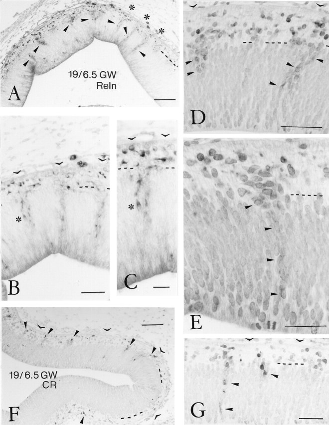
Reelin- and calretinin-ir neurons in the human preplate at stage 19, 6.5 GW. A, Low-power view of the rostral neocortex, immunostained for Reln. Arrowheadspoint to aggregates of Reln-positive cells in the VZ that seem to ascend to the preplate. Dashed line indicates border between VZ and preplate. Asterisks mark three radial columns of Reln-ir neurons in the VZ. Scale bar, 100 μm.B, Higher-power view of radial columns of Reln-ir neurons in the VZ. Open arrowheads indicate the pial surface, at places obscured by connective tissue. Scale bar, 50 μm.C, Higher magnification of the cell column marked by anasterisk in B and C, showing clearly the Reln immunoreactivity, which increases in intensity from the VZ to the preplate. Scale bar, 25 μm. D, Two Reln-ir cell aggregates (arrowheads) in the upper VZ, in continuity with Reln-positive neurons in the preplate. Scale bar, 80 μm. E, A single column of weakly Reln-ir cells (arrowheads) spans almost the entire width of the VZ. Scale bar, 40 μm. F, At stage 19, calretinin-ir (CR) neurons appear for the first time in the prospective neocortex, marking the beginning of the preplate period.Arrowheads point to aggregates of CR-ir neurons in the preplate. Scale bar, 100 μm. G, Occasionally, CR-ir cells are present in the VZ. Scale bar, 50 μm.