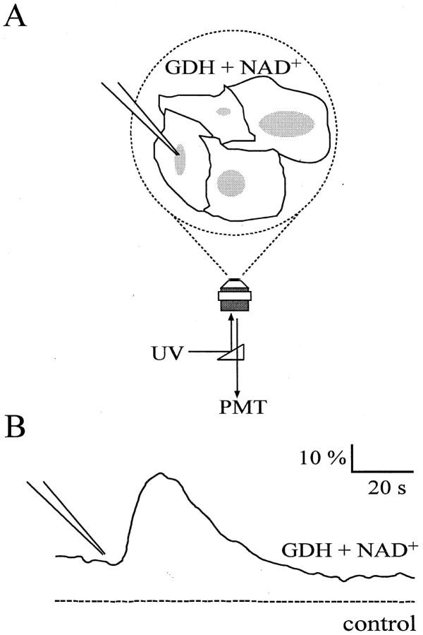
Fig. 2.
GDH can detect changes in extracellular glutamate levels that result from elevated internal calcium in astrocytes.A, Confluent cultures of purified astrocytes were bathed in a GDH saline. The entire optical field was illuminated by UV light, and the fluorescence emission was detected with a PMT. A glass pipette was used to gently tap the surface of an astrocyte to evoke a wave of elevated calcium and cause the release of glutamate. B, Fluorescence signals resulting from the accumulation of NADH that is generated as a result of the release of glutamate from stimulated astrocytes (continuous line). No changes in fluorescence were detected when GDH, NAD+, or both (dotted line) were omitted from the bathing solution. (Note that the signal shown by the dashed line is offset for display purposes.) The tip of the pipette in B indicates the time when pipette–cell contact occurred. Changes in fluorescence are expressed as dF/Fo(Fo = fluorescence level before cell stimulation; dF = change in fluorescence).

