
Fig. 3.
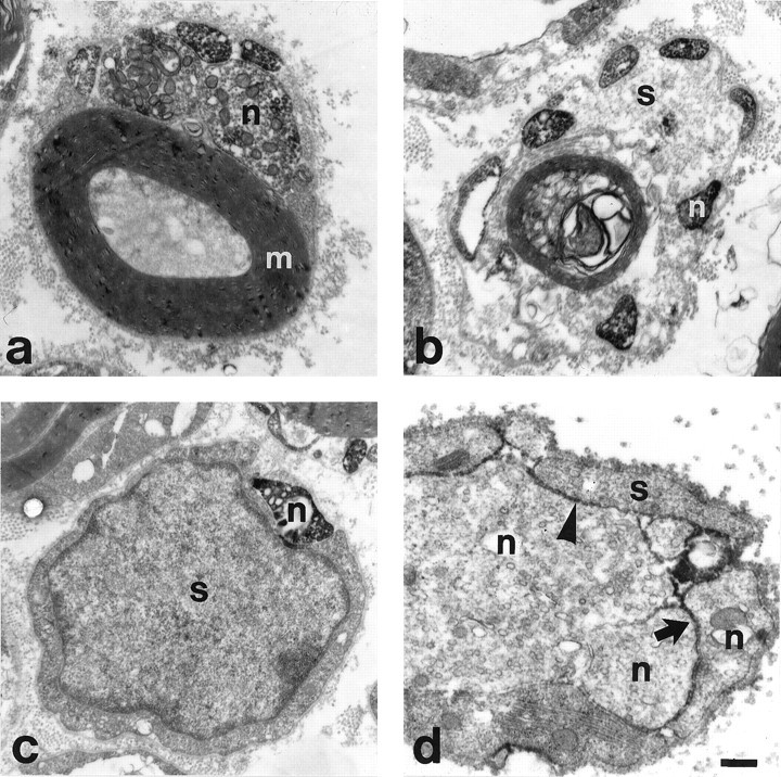
Regenerating facial neurites contain α7- and β1-integrin subunits at the ultrastructural level.a–c, α7- in regenerating motoneurites (n) 4 d after nerve crush in association with intact myelin (m) (a), with a partially demyelinated (b), and a completely demyelinated (c), α7-negative Schwann cell (s). The submembranous and cytoplasmic staining is attributable to immunoreactivity against the cytoplasmic part of the α7-subunit. d, The immune staining against the extracellular part of the β1-subunit is localized on the cell surface of regenerating neurites (n) and Schwann cells (s) at day 7. Pronounced staining at sites of axon–axon and axon–Schwann cell contacts (arrowhead). Scale bar, 0.45 μm.