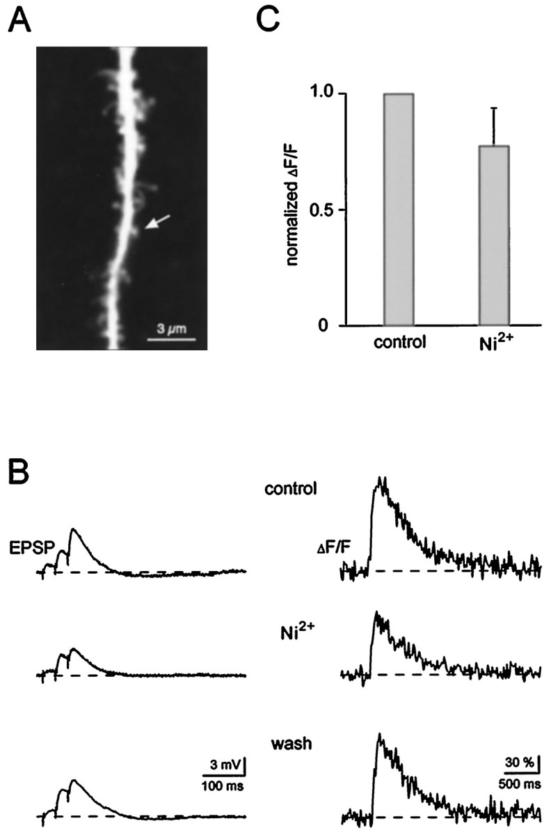
Fig. 3.

Low-threshold voltage-gated Ca2+ channels are not required for the generation of Ca2+ transients in spines. A,Confocal image of a dendritic segment containing an active spine (arrow). B, EPSPs (left traces) and associated spine Ca2+ signals (ΔF/F, right traces). Afferent fibers were stimulated with a short burst consisting of three stimuli given at 50 Hz. Application of nickel (40 μm), a blocker of T-type voltage-gated Ca2+ channels, reversibly reduced the spine Ca2+ signal only by ∼30%. Note that Ni2+ also slightly reduced the EPSP amplitude. The traces represent averages of three to five individual recordings. Membrane potential was −69 mV. C, Bar graph summarizing the effect of Ni2+ (40–50 μm) on the peak amplitude of the subthreshold Ca2+ responses (n = 10, mean + SD). In each of these experiments, five responses in control conditions and 10 min after wash in of Ni2+ were averaged.
