
Fig. 1.
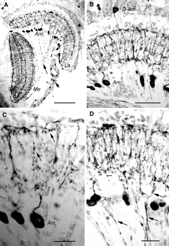
Tachykinin-related peptide in crayfish (P. leniusculus) optic lobe displayed by immunocytochemistry on cryostat sections (horizontal sections; peroxidase method). A, LomTK-immunoreactive (LTK-IR) neurons in theLa and the Me. Note the cell bodies ofAm cells below the lamina. Some LTK-IR axons are seen connecting the lamina and the medulla (longarrow). In the medulla four main layers of LTK-IR processes can be resolved. The cell bodies of the lamina–medulla neurons are seen at the shortarrow.B, Labeling with monoclonal antibody to substance P. TheLa in higher magnification with cell bodies ofAm and D-Am neurons is shown. Note the tangential processes running distally and proximally. C, Detail of substance P-immunoreactive Am neurons in the lamina. Note the varicose processes in the synaptic neuropil (arrows). D, LomTK-IR neurons in the lamina. Ams are seen together with lamina–medulla neurons (e.g., at arrows). Together they form superimposed tangential processes. Those of the lamina–medulla neurons are less prominently immunolabeled. Am, Amacrine;D-Am, displaced-amacrine; La, lamina;Me, medulla. Scale bars: A, 0.1 mm; B, 50 μm; C, D, 20 μm.