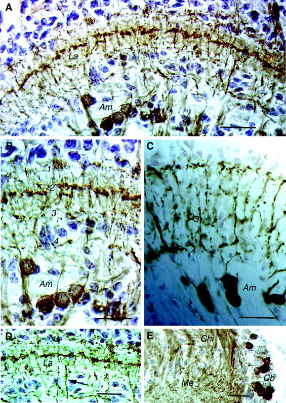
Fig. 2.

Cryostat sections of crayfish lamina (horizontal sections). A, GABA-immunoreactive neurons in the lamina of P. leniusculus (ABC method; counterstaining of nuclei with toluidine blue) are shown.Am neurons are labeled in addition to neurons connecting the La and the medulla (not clearly seen here; refer toD). Note the immunolabeled processes in three layers of the lamina; the midlayer of tangential processes is most prominent.B, Detail of the lamina with cell bodies of GABA-immunoreactive Ams and processes in the synaptic neuropil in four layers (1–4) is shown. The processes in layer 2 are derived from the lamina–medulla-connecting neurons. C, For comparison substance P-immunoreactive Ams (peroxidase method) are shown. Layers1, 3, and4 are indicated; no processes are seen in layer 2. D, GABA-IR processes of the neurons connecting the La and the medulla form a tangential layer in the midregion of the lamina neuropil. One of the lamina–medulla-interconnecting axons is seen at thearrow. E, Cb of lamina–medulla-interconnecting GABA-IR neurons reside adjacent to theMe neuropil close to the Chibetween the lamina and the medulla. Cb, Cell bodies;Chi, chiasm. Scale bars: A,D, E, 50 μm; B, C, 20 μm.
