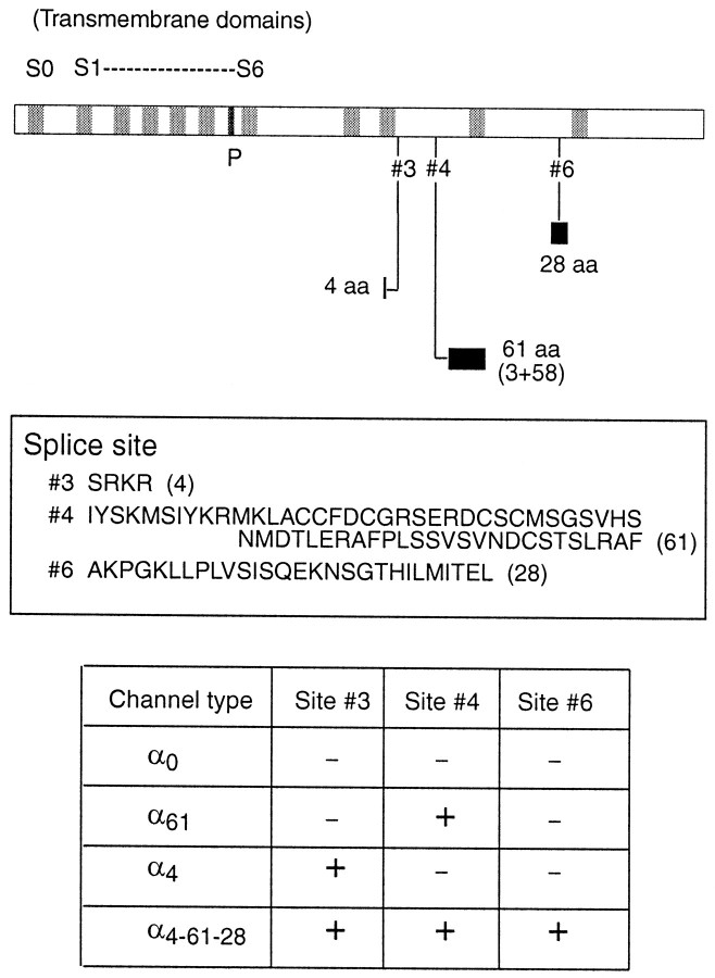
Fig. 1.
slo-α splice variants. A schematic layout of the slo-α gene is shown attop. The shaded areas are hydrophobic regions of the protein. The extended C-terminal region (afterS6) of the protein houses the calcium binding site and several splice sites (#3–#6). We have examined channels with a four amino acid insert at site #3(α4), a 61 amino acid insert at site#4 (α61), and a channel with both of these plus a 28 amino acid exon at site #6(α4–61-28), in addition to the full-length (α0) cloned initially. The amino acid sequences of the different alternative exons are shown in the middle panel. The bottom panel shows the channel types and the exon combinations present in each channel type. Splice sites are numbered according to Fettiplace and Fuchs (1999).

