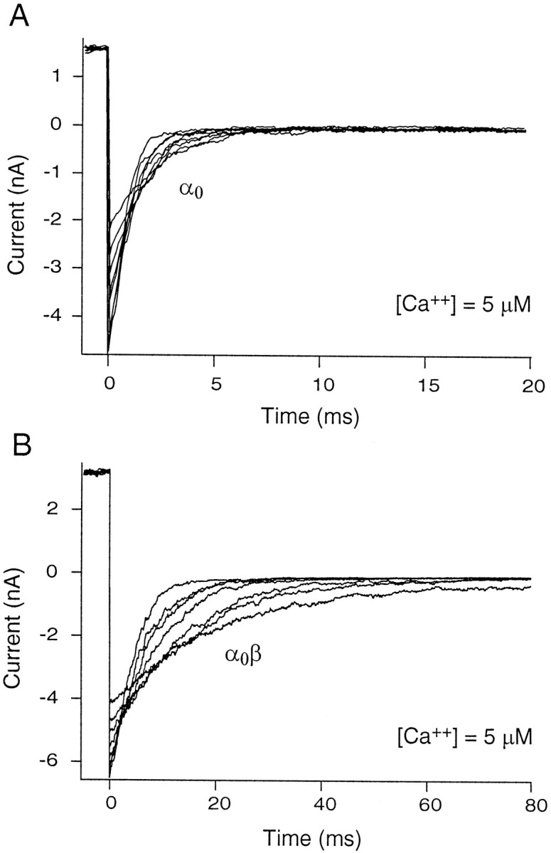
Fig. 6.

Effect of β subunit on deactivation time constant. A, Currents were measured from a macro patch containing α0. The channels were activated with a brief depolarization to +120 mV and then allowed to relax at a family of voltages ranging from −50 to −120 mV in steps of 10 mV. The panel shows decaying “tail” currents at a calcium concentration of 5 μm. The time constant of deactivation was determined by fitting single exponential functions of the form I= I0exp(−t/τ), whereI0 is the instantaneous current at the beginning of the deactivation pulse and τ is the time constant.B, Patches containing α0β were stepped to +60 mV and thereafter from −80 to −150 mV. Notice the substantial slowing of the tail currents (changed scale bar on x-axis) on β addition. Tail currents were slowed >10-fold by the addition of β subunits.
