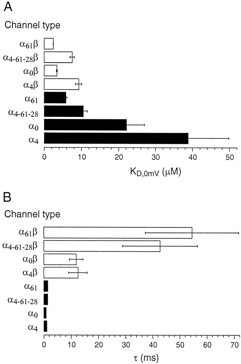
Fig. 8.

Calcium affinities at 0 mV (KD) and deactivation time constants were compared for the eight channel types created by four splice variants expressed either with or without β subunits.A, KD values for αx (filled bars) and αxβ (open bars) channels are shown, where x denotes the different exons.B, Deactivation time constants measured at −100 mV membrane potential and 5 μm “cytoplasmic” calcium for αx (filled bars) and αxβ (open bars) channels. The net range in deactivation kinetics between the fastest αx and the slowest αxβ is >50-fold. Error bars are standard errors from 5–10 experiments and are shown if they are larger than the width of the lines used in the bar plot.
