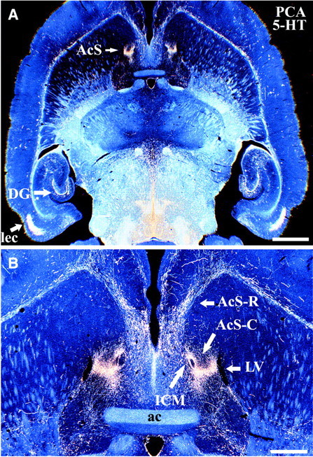
Fig. 5.

PCA-resistant 5-HT axons have a highly restricted localization in the caudal NAc shell. Low-magnification dark-field images show the distribution of drug-resistant varicose 5-HT-IR (SERT-negative) axon terminals in rat brain after PCA (2 × 10 mg/kg) treatment. Note the dense, restricted innervation by spared 5-HT-IR axons in the caudal NAc shell, as in dentate gyrus and entorhinal cortex (A). Higher magnification (B) reveals that the spared 5-HT axons in the caudal shell are situated between the lateral ventricle (laterally) and the island of Calleja magna (medially). Animals were killed 14 d after PCA treatment, and horizontal sections at the level of the NAc shell were processed for 5-HT immunocytochemistry. Scale bars:A, 2.0 mm; B, 600 μm.ac, Anterior commissure; AcS-C, caudal nucleus accumbens shell; AcS-R, rostral nucleus accumbens shell; DG, dentate gyrus; ICM, island of Calleja magna; lec, lateral entorhinal cortex;LV, lateral ventricle.
