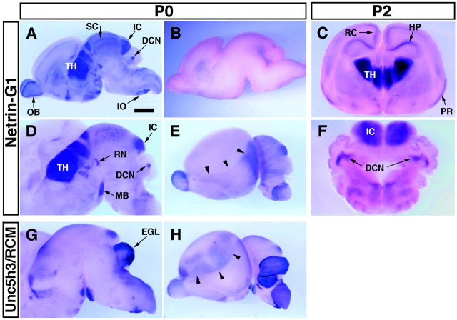
Fig. 6.
Regional distribution ofnetrin-G1 transcripts in mouse brain revealed byin situ hybridization. Parasagittal cuts of the P0 mouse brain (A, B, D,E, G, H) and P2 coronal vibratome slices of the P2 mouse brain (C,F) were hybridized with digoxigenin-labeled antisense cRNA probes specific for netrin-G1(A–F, except for B) orUnc5h3/rcm (G, H). Sense probe for netrin-G1 showed no signal under the conditions used (B). In the lateral views of cerebral hemisphere, distributions of netrin-G1(E) and Unc5h3/rcm(H) were complementary and delineated the boundary (arrowhead) between neocortex and allocortex. Scale bar: A, B, E,H, 1 mm ; C, 0.85 mm; F, 0.65 mm; D, G, 0.8 mm.DCN, Deep cerebellar nuclei; EGL, external germinal layer; HP, hippocampal formation;IC, inferior colliculus; IO, inferior olive; MB, mammillary body; OB, olfactory bulb; PR, piriform cortex; RN, red nucleus; RC, retrosplenial cortex; SC, superior colliculus; TH, thalamus.

