
Fig. 1.
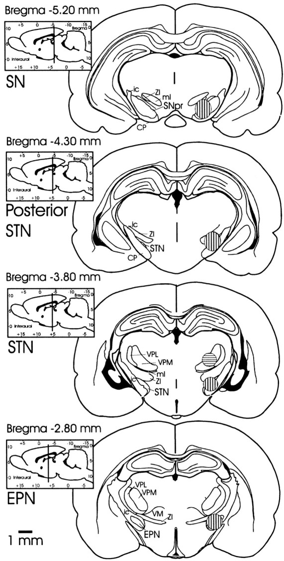
Coronal sections depicting areas of microinfusions into EPN, STN, dorsal to STN, posterior border of STN, and SNpr. Coronal sections for histology were sectioned in the plane corresponding to the atlas of Paxinos and Watson (1997) from which these drawings were derived. Striped areas (shown in one hemisphere only) designate the regions within which injection sites fell. Vertically stripedareas designate the regions of EPN, STN, posterior STN, and SNpr injection sites on respective coronal slices. The area of injection sites located dorsal to STN is shown on the same coronal slice as STN and is designated by the horizontallystripedarea. CP, Cerebral peduncle;EPN, entopeduncular nucleus; ic, internal capsule; ml, medial lemniscus; SNpr, substantia nigra pars reticulata; STN, subthalamic nucleus; VM, ventromedial nucleus of the thalamus;VPL, ventral posterolateral thalamic nucleus;VPM, ventral posteromedial thalamic nucleus;ZI, zona incerta.