
Fig. 3.
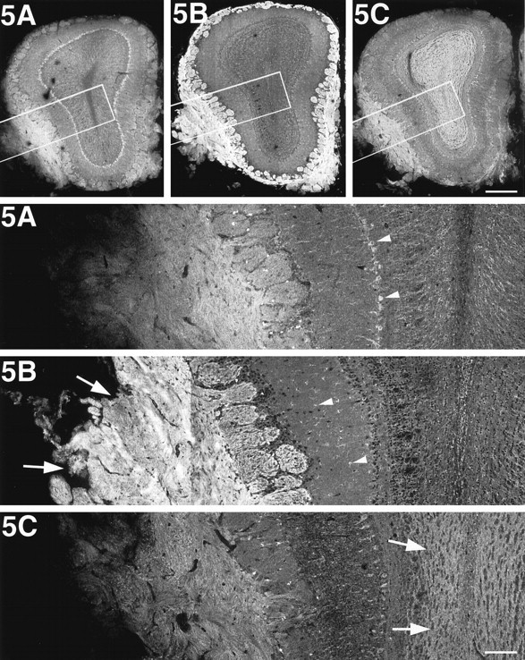
KIF5B is highly expressed in olfactory primary axons. Immunofluorescence of the olfactory bulb after staining with anti-KIF5 antibodies. The bottom panels represent high magnifications of the boxed areas of the top panels, respectively. KIF5B was highly expressed in the olfactory primary axons (arrows in 5B); however, glial cells, mainly astrocytes (arrowheads in5B), were stained predominantly in the rest of the olfactory bulb. On the other hand, KIF5A and KIF5C were localized only to the neuronal cells (e.g., mitral cell; arrowheads in5A). Note the strong staining of KIF5C in the granule cells (arrows in 5C). Scale bars:top, 0.4 mm (low magnifications); bottom, 0.1 mm (high magnifications).