
Fig. 7.
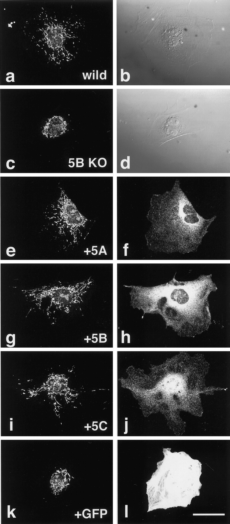
Rescue of abnormal mitochondrial localization in the KIF5B null mutant cells by each of the KIF5s.a, b, Wild-type control.c–l, KIF5B null mutant cells. Cells were injected with the expression vectors of KIF5A (e,f), KIF5B (g,h), KIF5C (i, j), or GFP (k, l). Mitochondrial staining using Mitotracker (a, c,e, g, i,k), Nomarski images (b,d), immunofluorescence after staining with each of the KIF5s (f, h, j), and fluorescence by exogenous GFP (l). Note the recovery of mitochondrial dispersion radiating from the cell center as in wild-type cells after microinjection of KIF5 but not after that of GFP plasmids. Scale bar, 50 μm.