Fig. 6.
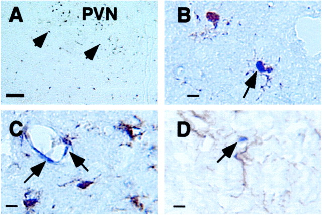
Representative bright-field microphotographs show labeling of IκBα mRNA in individual cells in the brain parenchyma of adrenalectomized animals killed at 2 hr after they received an intraperitoneal injection of 1 mg/kg LPS. A, Low-magnification microphotograph shows single-labeled IκBα mRNA-expressing cells (arrowheads) in the brain.B, Colocalization of IκBα mRNA expression and microglial immunostaining (arrow) is shown.C, Labeled IκBα mRNA-expressing cells (blue color) in endothelial cells (arrows; microglia were labeled by brown color) are shown.D, Labeled IκBα mRNA-expressing (arrow) cells do not colocalize with GFAP-stained (brown color) astrocytes. Scale bars: A, 50 μm; B–D, 10 μm.

