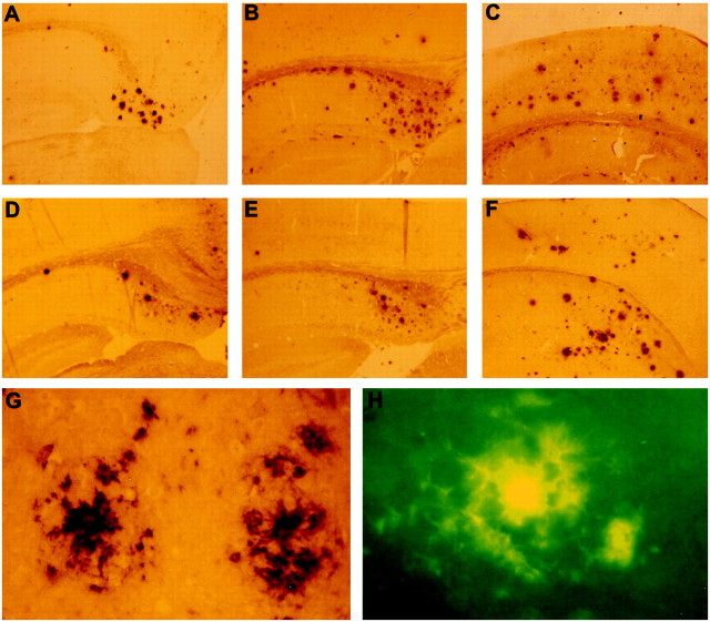
Fig. 2.
Immunohistochemical staining with antibody FCA18 of brain sections of single APP/V717I transgenic mice and double APP/V717I × PS1/A246E transgenic mice. Amyloid plaques in subiculum of double transgenic mouse at 8 months of age (A) and in subiculum (B) and cortex (C) at 13 months of age. Amyloid plaques in subiculum (D) of APP/V717I transgenic mouse (15 months) and in subiculum (E) and cortex (F) of APP/V717I transgenic mouse (18 months).G is higher magnification to illustrate the typical neuritic plaques with a dense core as described previously (Moechars et al., 1999a; Van Dorpe et al., 2000). H is an example of a typical thioflavine S-stained plaque, abundantly present in the brain of single and double transgenic mice (Van Dorpe et al., 2000).

