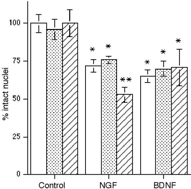
Fig. 5.
Blocking the actions of endogenously produced BDNF and/or NT4 with TrkB–IgG enhances neuronal loss in response to NGF. TrkB–IgG (10 μg/ml) was added to the cells 1 hr before the addition of neurotrophins for overnight treatment. Data are expressed as the percent of intact nuclei relative to that in the absence of added neurotrophins. Open bars indicate cells in the presence or absence of the indicated neurotrophins alone. Stippled bars show neurons with the IgG control fragment. Hatched bars indicate cells treated with TrkB–IgG. Anasterisk indicates values different from control atp < 0.001; double asterisksindicate a value different from NGF alone or the NGF + IgG control atp < 0.01. Data reported are from triplicate samples from two independent experiments (n = 6).

