
Fig. 1.
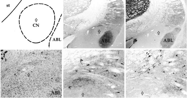
Schematic of the amygdala complex (top left) with the arrow indicating the position of the CN lesion at the level of the sections shown in the photomicrographs. The photomicrograph of a Nissl-stained section (bottom left) reveals heavy gliosis at the lesion site in the dorsal region of CN. Photomicrographs (center panels) show intact ChAT-immunopositive cholinergic neurons surrounding CN at two magnifications. Similarly, photomicrographs (right panels) show intact parvalbumin-immunopositive GABAergic neurons surrounding CN. ABL, Basolateral nucleus; st, striatum.