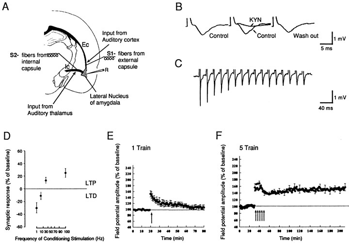
Fig. 1.
Monosynaptic field potential recording in the cortico-amygdala pathway. A, Schematic illustration of a coronal brain slice containing the amygdala. For the cortico-amygdala pathway a stimulating electrode was placed in the external capsule (S1); to stimulate the thalamo-amygdala pathway, we placed a stimulating electrode in the fibers from the internal capsule (S2). Field recordings were made from the lateral amygdala (R).B, The synaptic potential is blocked reversibly by kynurenic acid (KYN; 10 mm).Left, Before; middle, during KYN;right, 90 min after KYN was washed out.C, The synaptic potential recorded during a 50 Hz tetanus. The synaptic response followed the tetanus in a one-for-one manner without failure. D, LTD and LTP in the amygdala induced by different frequencies of stimulations. For 1–20 Hz, 900 pulses were applied; for 100 Hz, 100 pulses were applied. The changes in synaptic response were measured by the amplitude of field potential 15 min after the conditioning stimulus. E, E-LTP is induced by a single train of tetanus (100 Hz, 1 sec, indicated by thearrow). The LTP decayed to baseline in ∼40 min (n = 7; mean ± SEM). F, L-LTP is induced by five trains of tetanus (100 Hz, 1 sec at 3 min interval). The LTP is enduring and lasts at least 3 hr (n = 6; mean ± SEM).

